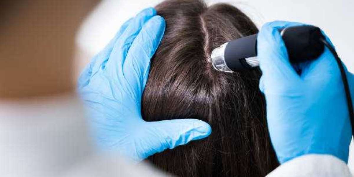

Hair thinning rarely affects every part of the scalp in exactly the same way. When considering Exosome Hair in Dubai, identifying where density has changed, where follicles remain active, and which areas are actually being treated can make the treatment plan more precise. Scalp mapping provides a structured way to document these differences instead of relying on a general description such as “overall thinning.”
Why Treatment Area Mapping Matters
A scalp can contain several different hair-loss patterns at the same time.
The frontal hairline may be receding while the crown remains relatively dense. Another person may have diffuse thinning across the top but comparatively stable sides and back.
Treating these situations as identical can lead to an unnecessarily broad or poorly defined treatment plan.
Mapping creates a clearer connection between the patient's actual hair-loss pattern and the proposed treatment area.
The Scalp Is Not One Uniform Surface
The scalp contains regions with different hair density, growth direction, follicular characteristics, and susceptibility to hair loss.
Common assessment zones include:
- Frontal hairline
- Temples
- Mid-scalp
- Central part
- Crown
- Upper sides
- Posterior scalp
- Donor region
Not every area necessarily requires the same level of attention.
The objective is to understand the distribution of the problem before deciding where an intervention may be appropriate.
Mapping Starts With Visual Assessment
The first step can be a systematic visual examination.
Rather than looking at the scalp as one surface, the clinician can compare different regions for changes in density, scalp visibility, hair caliber, and distribution.
Photographs can support this process when captured from consistent angles.
The value comes from consistency rather than simply collecting attractive images.
Why the Hairline Needs Separate Attention
The frontal hairline has considerable cosmetic importance because relatively small changes can alter the appearance of the entire face.
Hairline assessment can consider:
- Temple recession
- Frontal density
- Hairline shape
- Hair shaft thickness
- Presence of finer hairs
- Symmetry between sides
- Changes compared with previous photographs
A receding hairline does not automatically mean that the same strategy should be applied to the crown.
The biological and cosmetic objectives may be different.
Temple Thinning Can Be Easy to Miss
Temple recession can develop gradually.
Because the hairline changes slowly, a person may not recognize progression until older photographs are compared with current images.
The temples can also appear different depending on hairstyle and camera position.
Consistent documentation is therefore useful when determining whether the area is genuinely changing.
Crown Assessment Requires a Different View
The crown is particularly difficult to evaluate with a mirror.
A photograph from above can reveal scalp visibility that is difficult to observe from the front.
However, crown photographs are also highly sensitive to lighting and camera angle.
For that reason, repeated crown images should use similar positioning and illumination whenever possible.
Diffuse Thinning Needs Broader Mapping
Diffuse thinning does not necessarily produce one obvious treatment zone.
Instead, density may gradually decrease across the frontal, mid-scalp, and crown regions.
A person may describe this as “my whole top is getting thinner,” but mapping can reveal that certain areas are more affected than others.
This distinction can influence how a clinician defines the treatment objective.
Mapping Density and Mapping Hair Quality Are Different
A treatment map should not necessarily focus only on how many hairs are visible.
Hair caliber can also change.
Miniaturized hairs may be substantially finer than neighboring terminal hairs, creating a visual impression of reduced density even when follicles remain present.
Therefore, a useful assessment can consider both the number of hairs and the characteristics of those hairs.
Why Follicular Viability Matters
A visible thinning area does not automatically mean that follicles are absent.
Some regions may contain progressively miniaturized follicles that continue to produce fine hairs.
Other areas may have much more advanced loss.
This distinction is relevant because regenerative approaches are intended to influence existing biological processes rather than physically replace follicles in the way transplantation does.
Mapping Can Help Define Treatment Objectives
A treatment plan should ideally answer a simple question:
“What are we trying to improve?”
Possible objectives can include:
- Supporting existing hair density
- Addressing a specific thinning zone
- Improving the appearance of diffuse thinning
- Monitoring an area for future progression
- Supporting a broader hair-restoration strategy
The objective determines what should be measured afterward.
A Simple Scalp-Zone Framework
| Scalp region | What may be assessed | Why it matters |
|---|---|---|
| Hairline | Recession and frontal density | Highly visible cosmetic area |
| Temples | Symmetry and miniaturization | Can indicate progressive pattern changes |
| Mid-scalp | Overall density and shaft variation | Often important in diffuse thinning |
| Crown | Scalp visibility and density | Requires consistent overhead documentation |
| Sides | Baseline density | Useful for comparing affected and less-affected regions |
| Donor area | Density and follicular characteristics | Relevant when considering future restoration |
This is an assessment framework rather than a diagnostic staging system.
Mapping Before and After Treatment
The same regions should ideally be reviewed after treatment.
If the baseline assessment focuses on the crown but the follow-up photograph focuses mainly on the frontal hairline, the comparison becomes less useful.
Consistent regional assessment allows changes to be interpreted more carefully.
It also reduces the temptation to judge the treatment based on whichever area looks most favorable.
Why Standardized Photography Supports Mapping
Photographs can turn a subjective concern into a visual record.
Useful documentation may include standardized views of:
- Frontal hairline
- Left temple
- Right temple
- Mid-scalp
- Crown
- Left side
- Right side
The exact set can vary according to the patient's hair-loss pattern.
The important principle is to reproduce the same views during follow-up.
Trichoscopy Can Add More Detail
Ordinary photographs show the overall appearance of the scalp.
Trichoscopy provides magnified information that may help assess hair shaft variation, follicular openings, scalp surface characteristics, and other features.
This can be particularly useful when the visible difference between normal and miniaturized hairs is subtle.
Photography and trichoscopy therefore complement one another rather than serving exactly the same purpose.
Mapping Can Help Avoid Overgeneralization
A person may have significant thinning in one area but relatively stable hair elsewhere.
If the entire scalp is treated or described as equally affected without appropriate assessment, the treatment rationale becomes less precise.
A mapped approach allows the clinician to distinguish:
- Primary areas of concern
- Secondary areas to monitor
- Relatively stable regions
- Areas where another treatment may be more appropriate
This can improve the clarity of the overall hair-restoration strategy.
The Role of Existing Hair
Regenerative hair therapy is fundamentally different from follicular replacement.
A transplant moves viable follicles from a donor region to a recipient region.
A regenerative approach seeks to influence biological activity around existing tissue.
Consequently, areas containing existing but weakened or miniaturized hair may be assessed differently from areas with extensive long-standing absence of visible follicles.
Mapping Is Useful for Diffuse Hair Loss
Diffuse thinning presents a particular challenge because there may be no single bald spot to measure.
A structured map can identify where the reduction appears greatest.
This can also help distinguish a generalized change from isolated crown or hairline thinning.
The map becomes a reference point for future monitoring rather than simply a diagram of where injections might occur.
Dubai-Specific Factors in Scalp Mapping
The appearance of the scalp can change with environmental conditions.
Dubai's intense sunlight can increase scalp contrast in photographs, while sweating and humidity can cause hair strands to separate or clump.
Air-conditioning can also affect hair texture and styling.
These factors do not necessarily indicate biological changes.
When monitoring hair density in Dubai, consistent photography conditions can therefore be especially helpful.
What a Treatment Map Should Ideally Record
A useful treatment record may include:
- Date of assessment
- Areas of concern
- Baseline photographs
- Hair-loss pattern
- Scalp condition
- Relevant hair characteristics
- Treatment areas
- Planned follow-up intervals
- Outcome measurements
The level of documentation should be appropriate to the clinical setting.
The objective is to create enough information to evaluate whether the treatment plan remains logical over time.
Mapping Does Not Predict Treatment Success
A detailed scalp map improves assessment, but it cannot guarantee an outcome.
Hair growth depends on multiple biological factors, and response to regenerative interventions can vary.
Current clinical research on exosome-based hair treatment remains encouraging but limited. Systematic reviews have identified positive findings in several studies while emphasizing small samples, heterogeneous protocols, inconsistent outcome measures, and the need for stronger controlled research.
Mapping therefore supports better decision-making; it does not turn an emerging treatment into a predictable one.
Why Mapping Can Improve Patient Communication
Hair loss can be difficult to describe using words alone.
A patient may say, “My hair is getting thinner,” while a mapped assessment can show that the primary change is concentrated around the crown or mid-scalp.
This creates a more specific discussion between patient and clinician.
It also makes treatment goals easier to communicate and revisit.
Questions to Ask About the Treatment Area
Before beginning an exosome-based hair procedure, patients can ask:
- Which scalp areas are being treated?
- Why were these areas selected?
- Are different regions affected differently?
- Are there areas that should only be monitored?
- How will the treatment area be documented?
- Which outcome will be measured?
- Could another treatment be more appropriate for a particular region?
These questions can make the treatment plan easier to understand.
Mapping Creates a Better Baseline
The strongest reason to map the scalp is not to make the procedure appear more sophisticated.
It is to establish a meaningful baseline.
When the starting condition is clearly documented, future changes can be assessed against the same areas rather than against memory.
That is particularly useful when hair changes are gradual and difficult to perceive from day to day.
Turning a General Hair Concern Into a Structured Plan
Hair loss is rarely as simple as one number or one affected area.
The hairline, temples, mid-scalp, and crown can behave differently, and the visible appearance can be influenced by styling, lighting, and environmental conditions.
For Exosome Hair in Dubai, scalp mapping provides a practical framework for identifying where the concern exists, what characteristics should be measured, and how follow-up can be organized.
Patients exploring broader hair and aesthetic treatment options in Dubai can review the information available through Tajmeels Clinic before discussing their individual scalp pattern and treatment objectives with a qualified professional.
A precise treatment plan begins with a precise understanding of the area being treated.